

Occlusion for implant restorations depends on both clinical judgment and laboratory execution. Dental implants lack the periodontal-ligament-mediated mobility of natural teeth and have reduced proprioceptive feedback, so occlusal forces are transferred through a different biomechanical environment. For dentists and dental laboratories, the practical goal is to coordinate the bite record, opposing dentition, guidance, implant position, parafunctional risk, restorative design, and final clinical verification before small discrepancies become mechanical problems.

Why Occlusion for Implant Restorations Requires Different Thinking
Natural teeth move slightly within the periodontal ligament under load. That ligament also contributes to pressure perception and helps distribute functional forces. An osseointegrated implant is connected much more rigidly to bone, so the restoration does not benefit from the same mobility or sensory response.
This difference matters most when forces become heavy, lateral, repetitive, or poorly distributed. Mechanical and technical complications reported for implant restorations include ceramic chipping, screw loosening, abutment complications, loss of cement retention, and, less commonly, implant fracture.
Long-term data cited in the literature have reported ceramic chipping at about 20.31%, abutment and screw loosening at 5.3%, occlusal screw loosening at 2.57%, loss of cement retention at 2.06%, and implant fracture at roughly 0.5%. These numbers do not mean that occlusion is the sole cause of each complication. They do show why force management deserves attention throughout design, delivery, and maintenance.
The relationship between occlusal overload and mechanical complications is clearer than its direct relationship with peri-implant bone loss. Biological complications are multifactorial. Occlusion should therefore be treated as one important part of the overall risk environment, especially when the patient also has bruxism, long cantilevers, unfavorable implant positions, or previous prosthetic complications.
For the dental lab, this means implant occlusion cannot be reduced to making the crown "slightly light." Contact position, cusp form, opposing dentition, implant distribution, excursion, restorative material, and parafunctional risk all influence the final design. Many of the same record-related problems that create implant discrepancies also appear in conventional restorations, as discussed in our guide to high occlusion in dental crowns.
What the Dentist Must Communicate Before the Lab Designs the Occlusion
A digital scan gives the laboratory detailed anatomy. It does not automatically communicate every functional decision.
The laboratory still needs enough clinical information to understand how the restoration is expected to function in the patient's mouth.
Jaw Relation and Intended Occlusal Scheme
The starting point is a reliable jaw relationship. The dentist should provide an accurate bite record and indicate the intended reference position when that distinction is clinically relevant.
The laboratory also needs to know how the restoration should behave during function. Important information may include:
- Expected contact in closure;
- Anterior guidance;
- Canine guidance or group function;
- Lateral and protrusive movements;
- Whether the implant restoration should participate in excursion;
- Areas where posterior disclusion is expected.
Ambiguity in these areas forces the technician to infer the clinician's intent from static records. That may work in straightforward cases, but it becomes increasingly unreliable in complex implant rehabilitation.
Opposing Dentition and Functional Risk Factors
The antagonist is part of the restoration's functional environment. Natural enamel, zirconia, metal-ceramic restorations, an implant-supported prosthesis, and a removable denture do not behave identically under function.
The laboratory should know what the implant restoration opposes and, where relevant, what restorative material is present.
Bruxism and clenching also deserve explicit communication. A history of repeated ceramic fracture, screw loosening, heavy wear, or previous implant prosthetic complications can influence how conservatively the lab approaches cusp morphology, contact distribution, material selection, and excursive loading. In patients with active parafunction, clinicians may also consider a night guard for bruxism as part of the long-term protective strategy when clinically appropriate.
Implant Configuration and Provisional Feedback
Implant position and angulation define many of the restorative limits before CAD design begins. Useful information includes the implant system, platform, number of implants, implant distribution, angulation, restorative space, and any cantilever extension.
For complex anterior or full-arch cases, the provisional restoration can provide particularly valuable information. Wear facets, fracture, patient comfort, chewing efficiency, and observed excursive interferences show how the planned occlusal concept performs under real function.
A provisional that has worked well clinically gives the laboratory more than a shape to copy. It gives functional feedback that can guide the definitive restoration.
How the Dental Lab Translates Clinical Information Into Occlusal Design
Once the functional goals are clear, the laboratory converts them into restorative geometry.
Several design variables directly affect the way forces are introduced into the implant-supported restoration:
- Contact location;
- Contact intensity;
- Occlusal table width;
- Cusp height and inclination;
- Fossa width and depth;
- Surface morphology;
- Material thickness and support;
- Screw-access location where applicable.
A common design goal is to favor centrally positioned, controlled loading and reduce unnecessary lateral or off-axis force. In posterior implant restorations, this often means a broader fossa, less aggressive cusp inclines, and careful contact placement over a well-supported part of the restoration.
Axial loading is especially desirable when anatomy and implant position allow it. When the implant is angled, the restorative envelope is limited, or the screw-access channel compromises ideal morphology, the technician has to balance biomechanics, material support, and esthetics rather than follow a single formula.
Contact intensity also needs context.
In mixed tooth-and-implant dentitions, a graded contact approach is often appropriate. Natural teeth can contact earlier during light closure because of periodontal ligament resilience, while implant restorations participate more fully under firm closure. This is more useful than applying one universal micrometer target to every implant crown.
Dynamic occlusion deserves separate attention. Posterior implant restorations are generally protected from unnecessary working and non-working excursion contacts when natural teeth can provide guidance. If implants must participate in guidance, as can occur in extensive implant-supported rehabilitation, the entire occlusal scheme has to be considered rather than adjusting a single crown in isolation.

Occlusal Strategy Changes With the Type of Implant Restoration
There is no single occlusal prescription that fits every implant case. The design priorities change with the number of implants, restoration span, position in the arch, opposing dentition, and overall prosthetic concept.
Single Posterior Implant Crown
For a single posterior implant crown, the main goals are controlled static contact, favorable force direction, and minimal unnecessary excursive loading.
The laboratory should avoid overly steep cusps or small, concentrated contacts that create unfavorable force vectors. Where adjacent natural teeth can provide lateral guidance, the implant crown is usually kept out of unnecessary excursive interference.
The opposing arch matters. An implant crown opposing natural enamel is a different functional situation from one opposing another implant-supported restoration.
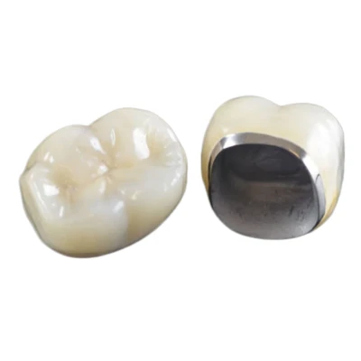
For readers comparing restorative options, our overview of implant crowns explains the main crown types and material considerations. For zirconia-specific cases, the zirconia implant crown page provides additional product-level information.
Anterior Implant Crown
Anterior implants require a different balance because esthetics and guidance are closely linked.
Palatal morphology, anterior coupling, centric freedom, and the patient's existing guidance pattern all need attention. If natural teeth can carry the initial guidance, that is often desirable. If the implant restoration must participate, the contact should be placed on a structurally well-supported part of the restoration and coordinated with the rest of the anterior segment.
An anterior implant crown can look excellent and still create trouble if the palatal design introduces a premature or heavy dynamic contact.
Multi-Unit Implant Bridge
Implant-supported bridges add force-distribution problems that are less important in a single crown.
The dentist and lab need to consider:
- Number and distribution of implants;
- Bridge span;
- Passive fit;
- Contact distribution;
- Connector and framework design;
- Cantilever presence and length.
Cantilevers deserve particular attention because they increase leverage on the supporting implants and prosthetic components. Their presence should be communicated clearly before occlusal design is finalized.
Passive fit is also important. A multi-unit implant restoration should not begin function with internal stress caused by an inaccurate framework and then receive additional occlusal loading on top of it.
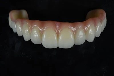
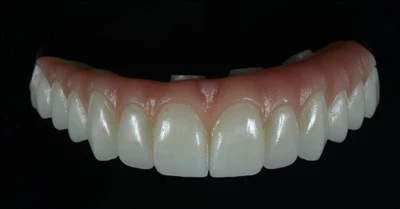
Full-Arch and All-on-X Restorations
Full-arch implant restorations require an arch-level occlusal strategy.
The technician cannot treat each unit as if it were an isolated implant crown. Implant distribution, distal cantilever, opposing arch, restorative material, anterior guidance, parafunction, and the provisional response all influence the final scheme.
This is also where provisional feedback becomes particularly valuable. If a provisional full-arch restoration shows repeated posterior wear, fracture, unstable contacts, or patient discomfort, those findings should be reviewed before the definitive prosthesis is manufactured.
There is no universal rule that every full-arch case should receive the same balanced, lingualized, or mutually protected scheme. The appropriate design depends on the prosthesis and its opposing arch. For case-specific product information, see our All-on-X implant bridge page.
How Digital Workflows Improve Dentist–Lab Occlusal Coordination
Digital workflows improve the transfer, visualization, and review of implant cases.
A complete digital implant case may include intraoral scans, an opposing arch scan, bite records, scan-body data, implant-library information, provisional scans, and additional functional records. For a broader explanation of what laboratories need from digital records, see what dental labs need from an intraoral scan.
CAD software then allows the technician to evaluate contact location, cusp morphology, restorative space, and virtual articulation before milling or printing begins.
Virtual articulators can also help assess lateral and protrusive movements when the required clinical records are available. The same principle applies to physical articulation: the device is only as useful as the records used to mount the case. Our article on dental articulators for predictable occlusion explains that role in more detail.
Their value still depends on the data supplied.
An inaccurate bite remains an inaccurate bite after it enters CAD software. Virtual articulation cannot identify unreported bruxism, previous screw loosening, a provisional fracture, or a clinician's intended guidance pattern unless that information reaches the laboratory.
For implant cases, digital case verification should therefore begin before design. Implant system and platform identification, scan-body and library compatibility, opposing records, and occlusal information should all be checked before manufacturing starts.
Digital technology improves repeatability. Complete communication gives that technology useful clinical meaning.
Common Dentist–Lab Coordination Errors That Create Occlusal Problems
Many delivery problems begin earlier in the workflow than the final milling or finishing stage.
A restoration that appears correct in CAD can still require significant chairside adjustment if the clinical records do not reproduce the patient's actual functional relationship.
|
Missing or Inaccurate Information |
Possible Clinical Result |
|
Inaccurate bite record |
Restoration delivers high, low, or uneven |
|
Incomplete opposing scan |
Incorrect contact relationship |
|
Missing excursion or guidance information |
Unexpected dynamic interference |
|
Unreported bruxism |
Functional risk is not reflected in the design |
|
Unknown opposing material |
Poor material or surface-design decision |
|
No provisional feedback |
A known functional problem may be repeated |
|
Incorrect scan-body or implant-library identification |
Fundamental restorative fit or design error |
Chairside adjustment does not automatically identify one single source of error. Seating differences, mandibular position, bite-record distortion, tooth movement, incomplete functional records, and true intraoral dynamics can all change the final contact pattern.
This is why a predictable implant workflow needs feedback in both directions. The dentist provides the clinical environment. The lab flags incomplete or conflicting information before manufacturing. When a complex design needs review, both sides should resolve the issue before the case reaches final production.
Delivery and Long-Term Occlusal Maintenance
Occlusal design is completed clinically.
The laboratory can design and verify the restoration against the records it receives, but the dentist must confirm the result intraorally at delivery.
Clinical verification should include seating, static contacts, excursive contacts, patient comfort, restoration stability, and the effect of any chairside adjustment. Adjusted ceramic or zirconia surfaces should be repolished appropriately so that the finished surface does not become unnecessarily abrasive to the opposing dentition.
Occlusion also changes with time.
Long-term studies have reported interproximal contact loss around implant restorations in roughly 29% of evaluated contact points, more often on the mesial side. Natural teeth can continue to move while osseointegrated implants remain comparatively fixed, and some patients develop infra-occlusion or changes in the relationship between an implant crown and adjacent teeth over time.
Annual maintenance should therefore include more than checking whether the implant is stable. A practical sequence is:
1.Review patient-reported changes, discomfort, food packing, clicking, fracture, or altered bite.
2.Check restoration retention, wear, chipping, screw access, and structural integrity.
3.Reassess static and dynamic occlusion, including signs of active bruxism and the condition of any protective splint.
4.Examine proximal contacts and positional changes relative to adjacent teeth and prostheses.
A restoration that functioned well at delivery can still require adjustment years later because the surrounding dentition and functional environment continue to change.
Dentist–Lab Checklist for Implant Occlusion
For routine implant cases, a short coordination checklist can prevent many avoidable assumptions.
|
Stage |
Key Information or Check |
|
Dentist → Lab |
Implant system/platform, accurate scans, opposing arch, bite relationship, intended occlusal scheme, guidance, bruxism/parafunction, cantilever, opposing material, provisional feedback |
|
Lab Review |
Scan-body/library compatibility, restorative space, implant angulation, contact distribution, cusp/fossa design, excursion, cantilever loading, material support |
|
Before Manufacturing |
Confirm unclear records, review complex designs, verify implant and occlusal parameters |
|
At Delivery |
Verify seating, static contacts, dynamic contacts, patient-specific function, stability, and polish after adjustment |
|
Maintenance |
Monitor screw retention, chipping, wear, proximal contacts, parafunction, splint condition, and occlusal changes |
The central principle is straightforward: implant occlusion works best when complete clinical information is translated into case-specific restorative design, then verified in the patient's mouth and maintained over time.
For dentists and dental laboratories working with an outsourced production partner, that coordination matters as much as manufacturing precision. ADS Dental Laboratory Ltd supports single implant crowns, implant bridges, All-on-X and other full-arch restorations through digital case receiving, CAD/CAM design, implant restoration workflows, occlusal inspection, and structured quality control. If you have a complex implant case or want to review the information your lab needs before production, contact ADS to discuss the case and workflow.