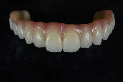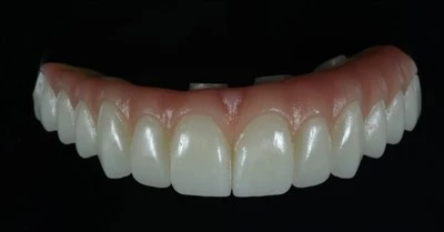
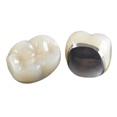
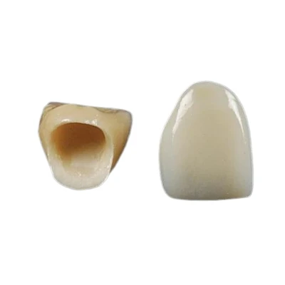
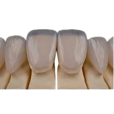
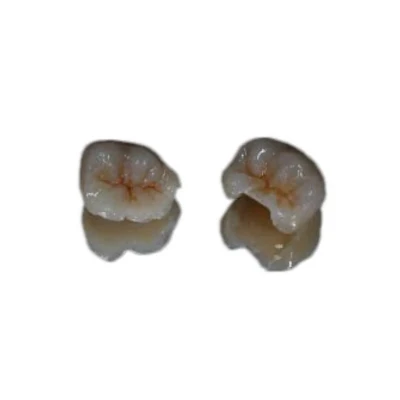

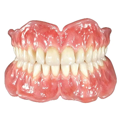
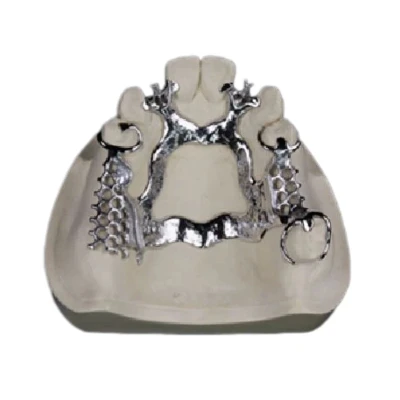
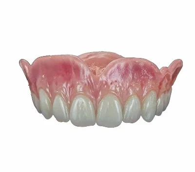

A digital denture is a complete or partial removable prosthesis designed and manufactured through a CAD/CAM workflow - intraoral scanning, computer-aided design, and subtractive milling or additive 3D printing - rather than conventional impression and flask-pack techniques.
The accuracy of a digital denture is not determined by any single machine, material, or software platform. It is the compounded result of how precisely each stage of the digital denture workflow captures, translates, and reproduces the patient's oral anatomy.
For dentists and dental laboratories, this means that digital denture accuracy, final fit, occlusion, retention, and the amount of chairside adjustment all depend on how well the complete workflow is controlled.

Five Stages That Determine Digital Denture Accuracy
Digital denture accuracy is a chain, and every link either preserves or degrades the dimensional truth of the original anatomy. The five stages are data acquisition, jaw relationship recording, CAD design, manufacturing method, and material behavior.
An error introduced at any stage propagates downstream - a 100-micron scanning inaccuracy becomes a 150-micron base discrepancy after design interpolation, and potentially larger after polymerization shrinkage during manufacturing.
What separates an accurate digital denture from an inaccurate one is how well each stage controls its own error budget and how tightly those stages are integrated.
For this reason, digital denture fabrication should be evaluated as a complete process rather than by looking only at the scanner, CAD software, milling machine, or 3D printer.
Intraoral Scanning and Digital Denture Accuracy
Trueness vs. Precision
Two metrics define scanner performance, and confusing them leads to wrong equipment choices.
Trueness measures how close the scanned data is to the actual physical anatomy - the systematic deviation from reality.
Precision, also called repeatability, measures how consistently the scanner produces the same result across multiple scans of the same object.
A scanner can be highly precise yet consistently inaccurate, producing the same wrong model every time.
For edentulous arches, trueness matters more than precision. The mucosa is mobile, compressible, and lacks fixed reference points like tooth surfaces. Edentulous arch scanning is therefore fundamentally more difficult than scanning a dentate arch with distinct tooth anatomy for stitch alignment.
The accuracy of this first step has a direct influence on subsequent denture base accuracy and tissue adaptation.
Scanner Technology and Its Limitations
Current intraoral scanners use optical principles such as confocal microscopy, triangulation, or optical coherence tomography. Each has trade-offs in how it handles the edentulous arch:
|
Scanner Principle |
Strength |
Limitation for Edentulous Arches |
|
Confocal optical scanning |
High trueness on smooth surfaces; handles wet fields well |
Stitching errors on long, featureless spans |
|
Triangulation-based scanning |
Fast capture; effective on clearly defined surface anatomy |
Can struggle with glossy mucosa and deep vestibules |
|
Optical coherence tomography |
Depth-resolved data; minimal powder needed |
Slower; limited clinical validation for full-arch dentures |
The edentulous arch lacks the anatomical landmarks that help stitching algorithms align consecutive image frames. A 2024 expert consensus on intraoral scanning noted that scanning strategy - the path the operator follows - directly affects accuracy, and that no unified standard yet exists for edentulous arch scanning protocols.
Scanning Strategy Matters
For edentulous patients, the scanning trajectory is not a minor technical detail - it is a determinant of digital denture fit.
The recommended approach scans the crest first, then extends laterally into the vestibular and lingual sulci. Scanning the vestibule under functional tension, with the patient moving the cheeks and tongue, captures the dynamic border that determines denture seal.
A static scan of the resting mucosa may produce a denture base that fits the anatomy at rest but loses seal under function.
Clinical studies show that as the edentulous span increases, both trueness and linear accuracy of digital impressions decline significantly. Kennedy Class I arches with free-end extensions show measurably lower accuracy than bounded defects.
For laboratories receiving digital cases, understanding what is required from an intraoral scan for predictable restorations can help reduce downstream errors before CAD design begins.
- The scan is the foundation. No amount of sophisticated digital denture design or manufacturing can recover accuracy that was never captured.

Jaw Relationship Records and Denture Occlusion
The Centric Relation Problem
Recording the correct vertical dimension of occlusion (VDO) and centric relation (CR) is where many digital dentures fail - not necessarily in the denture base fit, but in the occlusion.
A perfectly adapted base with the wrong jaw relationship is still an inaccurate denture.
Digital workflows offer two approaches:
- Digital gothic arch tracing: Electronic jaw recording devices capture CR in three dimensions, feeding data directly into the CAD software. This reduces transfer error but requires additional hardware and patient cooperation.
- Scan-based bite registration: A pre-existing denture or wax rim is scanned in occlusion, and the spatial relationship is extracted from the scan. This is faster but inherits any error in the rim or existing prosthesis.
Why This Stage Fails
The most common failure is not necessarily the recording device. It is the assumption that a single static record represents the patient's functional jaw position.
Patients with resorbed ridges and unstable neuromuscular patterns may show 0.5–1 mm of discrepancy between a recorded CR and their habitual closure path. That discrepancy translates directly into occlusal interferences on the finished denture.
A digital workflow does not eliminate this problem. It can simply make it faster to manufacture a denture from inaccurate jaw relationship data.
For this reason, digital denture accuracy should always include occlusal accuracy, not only tissue-side adaptation.
CAD Design Parameters That Affect Denture Fit
Design Parameters That Affect Accuracy
The digital denture CAD design stage is where raw scan data becomes a physical prosthetic design. Key parameters that determine final accuracy include:
- Tissue stop placement: Where the software places rigid contact zones on the cast affects how the base seats. Poorly placed tissue stops can cause rocking.
- Post-dam design: The posterior palatal seal depth and width determine retention. Software-generated post-dams that are too shallow may produce loose dentures, while excessive depth may cause tissue trauma and base lift.
- Border extension: The digital border must match the functional vestibule, not simply the resting vestibule. Over-extension can cause dislodgement; under-extension compromises seal.
- Tooth arrangement: Occlusal plane inclination, tooth positioning relative to the ridge crest, and cusp inclination all affect stability. A 2026 study demonstrated that measuring the intercrestal line angle and customizing tooth arrangement accordingly improved denture stability significantly over software-default arrangement.
These parameters show why CAD/CAM denture accuracy depends on both software capabilities and the clinical and technical judgment applied during design.
Software Is Not Neutral
Different CAD platforms can handle the same scan data differently.
The interpolation algorithms that fill gaps in scan data, default settings for base thickness, and automatic tooth placement logic can all vary. A denture designed in one software environment and manufactured on one machine is not automatically interchangeable with the same data processed in another system without re-validation.
The design stage is also where technician judgment remains especially important.
Software can automate geometry. It does not automate clinical judgment.
Milling vs. 3D Printing: How Manufacturing Affects Accuracy
Subtractive Manufacturing: Milled Dentures
Milled digital dentures are produced from pre-polymerized PMMA pucks.
Because the material is already polymerized under controlled industrial conditions, there is no polymerization shrinkage during the milling stage itself. This is one of the most important accuracy advantages of milling.
|
Parameter |
Milled PMMA |
Printed Resin |
|
Polymerization shrinkage |
None during milling from a pre-polymerized puck |
0.1–0.3% dimensional change |
|
Flexural strength |
65–90 MPa (meets ISO 20795) |
50–65 MPa (below threshold for some applications) |
|
Dimensional stability over 14 days |
Stable |
Measurable warping, greatest at posterior palatal seal and tuberosity regions |
|
Surface finish |
Dense, polishable |
Layer lines require post-processing |
|
Equipment cost |
High, particularly for 5-axis milling |
Lower for many DLP/SLA printing systems |
A 2024 study quantified that DLP-printed maxillary denture bases showed measurable deformation over 14 days, with the greatest warping occurring at the posterior palatal seal region and bilateral tuberosity areas - precisely the zones most critical for retention.
This is not simply a theoretical concern. It represents a measurable change in the denture base fit that was originally designed.
Additive Manufacturing: 3D-Printed Dentures
3D-printed dentures offer speed, cost efficiency, and the ability to produce complex geometries.
However, several variables influence 3D-printed denture accuracy, including:
- Print orientation
- Layer thickness
- Resin properties
- Support placement
- Washing procedures
- Post-curing protocol
Printed resins undergo light-curing that introduces dimensional change. A systematic review of digital removable denture manufacturing found that subtractive manufacturing demonstrated superior occlusal accuracy compared with additive manufacturing and conventional methods.
The Practical Verdict
Milling remains the accuracy benchmark for definitive digital dentures in the context discussed here.
Printing is viable for try-in prostheses, surgical guides, and provisional dentures where sub-100-micron precision may not be the primary requirement.
For a definitive prosthesis where long-term fit and retention matter, the dimensional stability of pre-polymerized milled PMMA provides an important manufacturing advantage.
This does not mean that every milled denture will automatically fit better than every printed denture. Equipment calibration, CAD design, material selection, manufacturing protocols, post-processing, and digital denture quality control still determine the final clinical result.
Material Behavior and Dimensional Stability
PMMA: Still the Standard
Polymethyl methacrylate (PMMA) has been used as a denture base material for over 80 years because of its combination of dimensional stability, mechanical strength, repairability, and biocompatibility. It also remains a widely used base material for an acrylic full denture.
CAD/CAM PMMA pucks are polymerized under high pressure and temperature in industrial settings, producing a material with significantly lower residual monomer content and higher flexural strength than hand-processed PMMA.
A 2026 comparative study of four digital denture base resins found that milled CAD/CAM PMMA and milled ADT resin both met the flexural strength requirement for complete denture bases above 65 MPa, while two 3D-printed resins fell below this threshold.
When Material Choice Undermines Accuracy
Even the best scan and digital denture design cannot compensate for a material that deforms in service.
Printed resins that absorb water, creep under occlusal load, or warp during post-cure can cause a denture that fit accurately at delivery to become loose or uncomfortable over time.
The initial accuracy may have been real. The material failed to maintain it.
This distinction between manufacturing accuracy and long-term dimensional stability is important when evaluating a digital denture material.
How Errors Accumulate Across the Digital Denture Workflow
Accuracy is not simply additive - errors can interact and compound throughout the digital denture workflow.
A scan with 50 microns of trueness error, a design that introduces 30 microns of interpolation deviation, and a manufacturing process with 100 microns of shrinkage do not necessarily produce a denture with exactly 180 microns of total error. The final base may deviate 200–300 microns from the intended fit because each stage influences the next.
The table below summarizes how each stage contributes to or controls the total error budget:
|
Stage |
Primary Error Source |
Typical Magnitude |
Mitigation |
|
Intraoral scan |
Stitching on featureless mucosa |
50–200 microns |
Functional border scanning; experienced operator |
|
Jaw relationship |
Static CR vs. habitual closure |
0.5–1 mm occlusal discrepancy |
Gothic arch tracing; try-in verification |
|
CAD design |
Default border and post-dam settings |
100–300 microns |
Customized parameters; technician judgment |
|
Manufacturing |
Polymerization shrinkage in printing |
0.1–0.3% linear |
Milled PMMA for definitive prostheses |
|
Material aging |
Water sorption, creep |
Progressive over months |
High-grade PMMA; proper post-cure for printed resins |
This is why evaluating only the nominal accuracy of a scanner, printer, or milling machine gives an incomplete picture of digital denture manufacturing accuracy.

What Actually Determines Digital Denture Accuracy
The accuracy of a digital denture is determined by the weakest link in the workflow - not by the most advanced piece of equipment.
A five-axis mill cannot fix a poor scan. A highly accurate scanner cannot compensate for incorrect jaw relationship data. Sophisticated CAD software cannot completely overcome material shrinkage in a printed base.
The practices that consistently produce accurate digital dentures share three traits: they scan with a strategy designed for edentulous arches, they verify the jaw relationship with a try-in before final manufacturing, and they select a manufacturing method and material appropriate for the required level of dimensional stability.
Everything else is optimization around those fundamentals.
A 2025 systematic review and meta-analysis of 19 randomized controlled trials involving 498 patients found that while digital and conventional dentures showed comparable clinical outcomes in retention, masticatory force, and patient satisfaction, digital workflows significantly reduced impression time and the number of post-insertion adjustment points.
The accuracy advantage of digital dentures is real - but only when every stage of the workflow is executed with discipline.
Conclusion
What determines the accuracy of a digital denture is not one scanner, one CAD program, one milling machine, or one material. Digital denture accuracy is the cumulative result of accurate clinical data acquisition, reliable jaw relationship records, appropriate CAD design, controlled manufacturing, stable materials, careful post-processing, and consistent quality control.
For dentists and dental laboratories, the most reliable digital denture workflow is therefore one in which every stage is controlled and verified rather than relying on equipment specifications alone.
ADS Dental Laboratory Ltd is a full-service digital dental laboratory in Shenzhen, China, providing long-term dental lab outsourcing and customized restorative production for overseas dentists and dental laboratories. Our capabilities include digital denture design, CAD/CAM processing, digital scanning, milling, 3D printing, case tracking, and structured quality control. Contact us to discuss your digital denture cases, production requirements, or long-term outsourcing needs.
FAQ
How accurate are digital dentures?
Digital dentures can achieve a high level of dimensional accuracy, but the final result depends on the entire workflow. Intraoral scan quality, jaw relationship records, CAD design, milling or 3D printing parameters, material stability, and post-processing can all influence the final denture fit.
A high-accuracy manufacturing machine cannot compensate for inaccurate clinical data captured earlier in the workflow.
What causes a digital denture to fit poorly?
Poor digital denture fit may result from inaccurate edentulous arch scanning, incomplete border capture, incorrect jaw relationship records, inappropriate CAD parameters, manufacturing distortion, polymerization shrinkage, or errors during post-processing.
Identifying the source requires evaluating the entire digital workflow rather than assuming the problem originates in a single production step.
Are milled dentures more accurate than 3D-printed dentures?
Milled dentures made from pre-polymerized PMMA generally offer strong dimensional stability because the material does not undergo polymerization shrinkage during the milling process.
3D-printed dentures can also achieve clinically useful accuracy, but print orientation, resin properties, layer thickness, washing, and post-curing can introduce dimensional variation.
The final result therefore depends on both the manufacturing method and how well the process is controlled.
Does intraoral scanning affect digital denture accuracy?
Yes. Intraoral scanning is one of the most important factors affecting digital denture accuracy, particularly in completely edentulous arches.
Featureless and mobile mucosal surfaces make image stitching more difficult than scanning dentate arches. Scan strategy, border capture, operator technique, and the length of the scanned span can all affect the accuracy of the resulting digital model.
How can a dental laboratory improve digital denture consistency?
A dental laboratory can improve consistency by standardizing case receiving, scan evaluation, CAD design parameters, jaw relationship verification, manufacturing settings, post-processing, and final quality control.
Equipment calibration, technician traceability, repeatable production protocols, and structured inspection are especially important when a laboratory handles digital denture cases at scale.